Ultherapy Before-and-After Photos: How to Judge What They Show
Written by Ultherapy in Seoul editorial team · PublishedAn Ultherapy before-and-after pair shows one person in two photographs. Standardized photographs help assess visible change in the person shown. They do not establish your likely result; interpreting study outcomes also requires information about patient selection, other treatments and follow-up. In studies where masked raters compared standardized photographs, measured changes were small, such as an average brow elevation of 1.7 mm at day 90[1], and in several studies blinded improvement rates were lower than patients' or physicians' own ratings[2,3,4]. Check the conditions of the two photographs before reading the difference between them as a result.

What one photo pair can show
If the photographs are comparable, a pair can show that one person's appearance changed between two moments. It cannot show how often a similar change happens, how large it is for other people or how long it lasts. Estimating frequency and duration requires group data and repeated follow-up.
The US Federal Trade Commission's staff Health Products Compliance Guidance (December 2022) draws the same line. It says individual consumer experiences are never sufficient to substantiate claims about a health product's effects, and that testimonials reporting results more dramatic than users can generally expect are likely to be deceptive. One worked example, an ad for a weight-loss supplement, pairs before-and-after photographs with a customer's accurate account of her result; the guidance finds that a fine-print disclaimer does not tell readers that their own result is likely to be much smaller.
What to check in a before-and-after set
Technical guidelines for applying the International Skin Imaging Collaboration's dermatological imaging standards list the conditions to standardize before skin images can be compared: lighting, background color, field of view, image orientation, focus and depth of field, resolution, and scale and color calibration[5]. They were written for dermatology imaging in general rather than for galleries.
- Matching conditions. Check that lighting, camera position, expression, posture and processing match[5].
- Selection and blinding. Ask who selected the photographs and what information assessors were blinded to.
- Follow-up day. The second photo should state the day after treatment. One rater-blinded study took standardized front and side photographs at days 2, 7, 28, 60 and 90[1]. That study reported mild, short-lived redness and swelling as common, so a photo from the first days may show swelling rather than the gradual change. Recovery is described under aftercare and downtime.
- Number of patients. Ask how many patients with a similar starting point were treated and how many photo sets exist.
What masked-rater studies found
The studies below report photographic assessments by masked, or blinded, raters alongside other ratings.

- In a 2010 rater-blinded cohort study of an investigational focused ultrasound device, three physician raters who were not study authors compared photographs taken before treatment and at day 90. They judged 30 of 35 participants (86%) to show a clinically significant brow lift; the average measured brow elevation was 1.7 mm. The authors could not quantitatively measure lower-face tightening because that area lacks fixed anatomical landmarks[1].
- In a retrospective study of women treated with MFU-V, physicians rated 77.7% as improved at 180 days on a global aesthetic scale, while blinded reviewers judged 67% to show improvement in appearance[2].
- In a 20-person MFU-V pilot, 95% were rated improved on physician and patient global aesthetic scales at one year, while blinded assessment rated 6 of 14 evaluable participants as improved at that point[3].
- Two blinded dermatologists comparing paired lower-face photographs scored 20.9% as improved, 62.5% as unchanged and 16.7% as worse, while 45.9% of patients reported improvement. A 24-patient study using Microson, a different ultrasound device, reported these ratings at a median 4.3 months[4,6]. These are not Ultherapy-specific outcome rates.
- A 2023 systematic review of 16 studies with predominantly female participants, using several ultrasound devices, reported measured brow lifts of 0.47–1.7 mm and submental reductions of 26–45 mm² on side-view photographs, and called for more objective outcome measures[6].
These studies reported small measured changes and differences between photographic and participant ratings.
What two FDA records show about photographs
Two of the Ulthera System's US 510(k) records include applicant-written summaries of the clinical data behind an added indication. Both relied on photographs.
The K121700 summary (cleared October 2, 2012) covers lifting lax submental and neck tissue. It reports that 51 of 70 patients reached a measured lift of at least 20 mm², and that masked evaluators identified improvement in 84.3% of those 51.
The K134032 summary (cleared June 20, 2014) covers lines and wrinkles of the décolletage. It states that the planned wrinkle scale could not be validated because its intra-rater and inter-rater reproducibility were low. The primary endpoint was changed to a post-hoc masked assessment of before and after photographs, with no success criteria set in advance. Of 108 per-protocol participants, 54 day-180 photo sets were set aside for inconsistencies including changes in lighting, color, focus, patient positioning and cropping. In the remaining 54, masked assessment recorded 36 (about 67%) as improved, 13 (24%) as "incorrect" and 5 (9%) as no change; the summary does not define the "incorrect" category further.
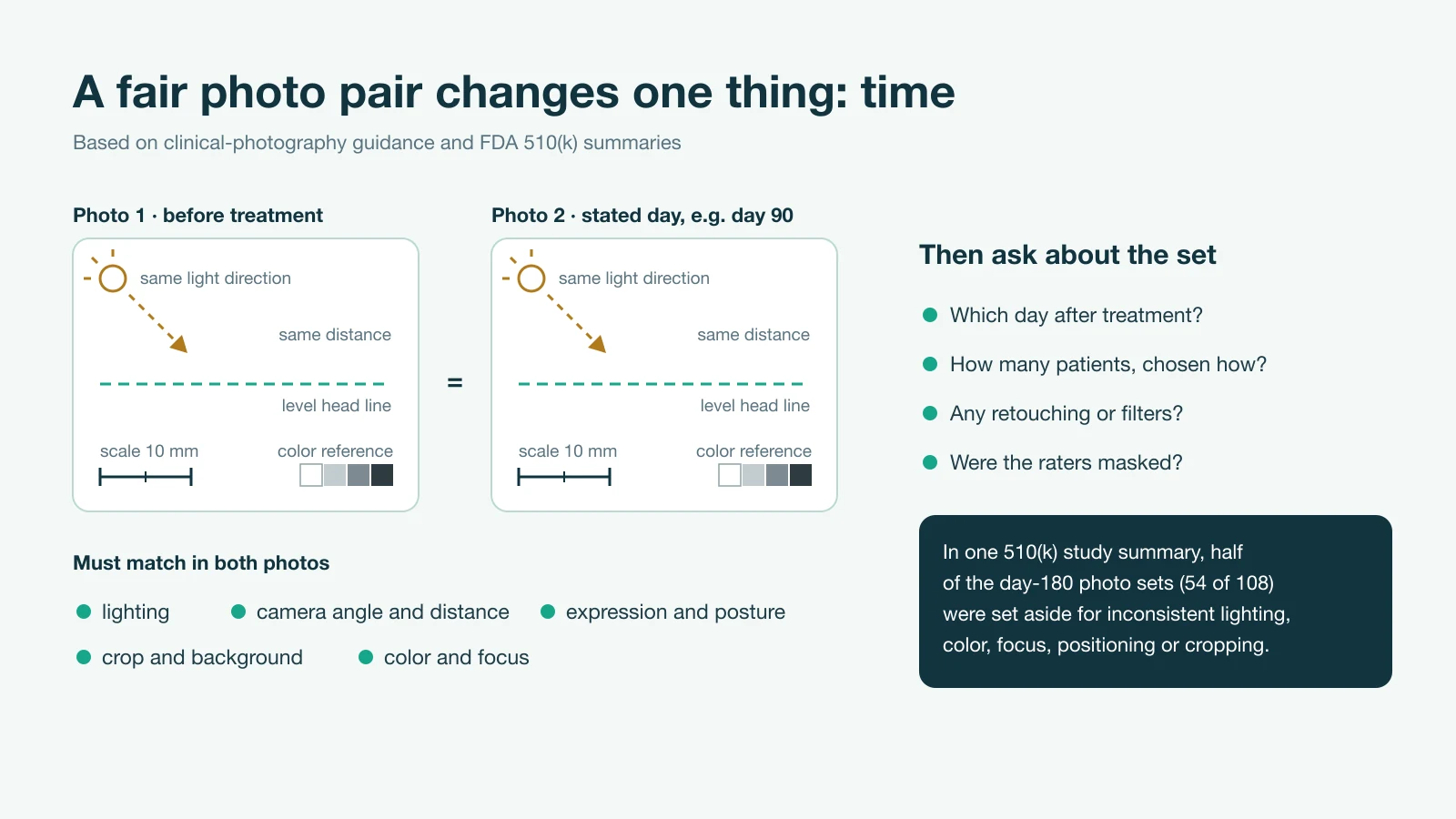
A gallery that does not say how its photographs were taken gives no way to know whether they would have passed that screen. The wider US regulatory history is covered in what Ultherapy is.
Photos in Korean medical advertising
Article 56 of Korea's Medical Service Act lists kinds of medical advertising that medical providers may not run. They include advertising, such as patients' accounts of their treatment, that may lead consumers to misunderstand treatment effects; advertising that leaves out important information such as serious side effects; and advertising that exaggerates objective facts.
Questions to ask about a clinic's photos
- On what day after treatment was the second photo taken?
- Were both photos taken with the same lighting, camera distance and pose?
- Has either image been filtered, retouched or cropped differently?
- How many patients with a starting point like mine have photo sets, and how was this one chosen?
- Who judged the change, and did they know which photo came first?
- Will you photograph me the same way at baseline and at follow-up?
For comparison, Delight Dermatology Clinic’s before-and-after section shows how one clinic presents its result photographs.
Common questions
Are Ultherapy before-and-after photos proof that the treatment works?
When is an Ultherapy after photo meaningful?
Why might photos show less change than I feel?
References
- Alam M, White LE, Martin N, et al. Ultrasound tightening of facial and neck skin: a rater-blinded prospective cohort study. J Am Acad Dermatol. 2010;62(2):262-9.
- Fabi SG, Goldman MP. Retrospective evaluation of micro-focused ultrasound for lifting and tightening the face and neck. Dermatol Surg. 2014;40(5):569-75.
- Werschler WP, Werschler PS. Long-term Efficacy of Micro-focused Ultrasound with Visualization for Lifting and Tightening Lax Facial and Neck Skin Using a Customized Vectoring Treatment Method. J Clin Aesthet Dermatol. 2016;9(2):27-33.
- Yalici-Armagan B, Elcin G. Evaluation of microfocused ultrasound for improving skin laxity in the lower face: A retrospective study. Dermatol Ther. 2020;33(6):e14132.
- Finnane A, Curiel-Lewandrowski C, Wimberley G, et al. Proposed Technical Guidelines for the Acquisition of Clinical Images of Skin-Related Conditions. JAMA Dermatol. 2017;153(5):453-457.
- Contini M, Hollander MHJ, Vissink A, Schepers RH, Jansma J, Schortinghuis J. A Systematic Review of the Efficacy of Microfocused Ultrasound for Facial Skin Tightening. Int J Environ Res Public Health. 2023;20(2):1522.