What Ultrasound Imaging Shows During Ultherapy
Written by Ultherapy in Seoul editorial team · PublishedUltherapy combines ultrasound imaging with focused ultrasound treatment. In the current US instructions, the DeepSEE display shows tissue from the surface to 8 mm, while the listed treatment transducers focus at 1.5, 3.0 or 4.5 mm. The image helps the trained user check skin contact and an appropriate treatment depth. It is not documented as a test that predicts an individual's final appearance.
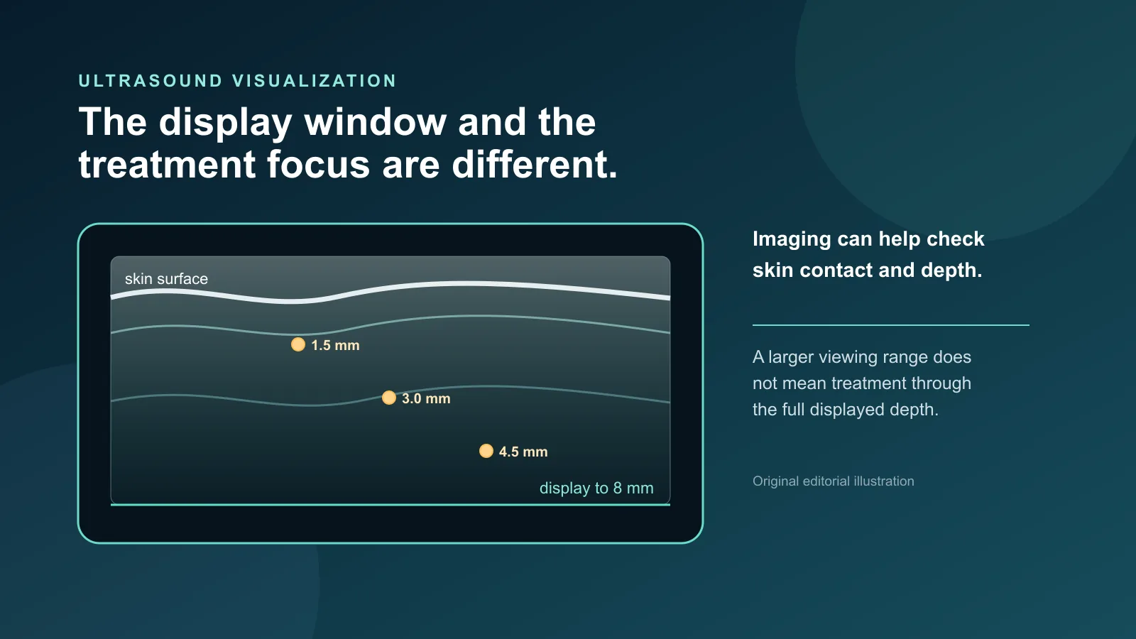
The phrase "ultrasound visualization" can make the screen sound like a diagnostic scan or an outcome forecast. The public device records describe a narrower role. They say the operator may visualize the tissue in the treatment path, confirm coupling between the transducer and skin, and check the selected treatment depth.
The imaging window and treatment depth are different
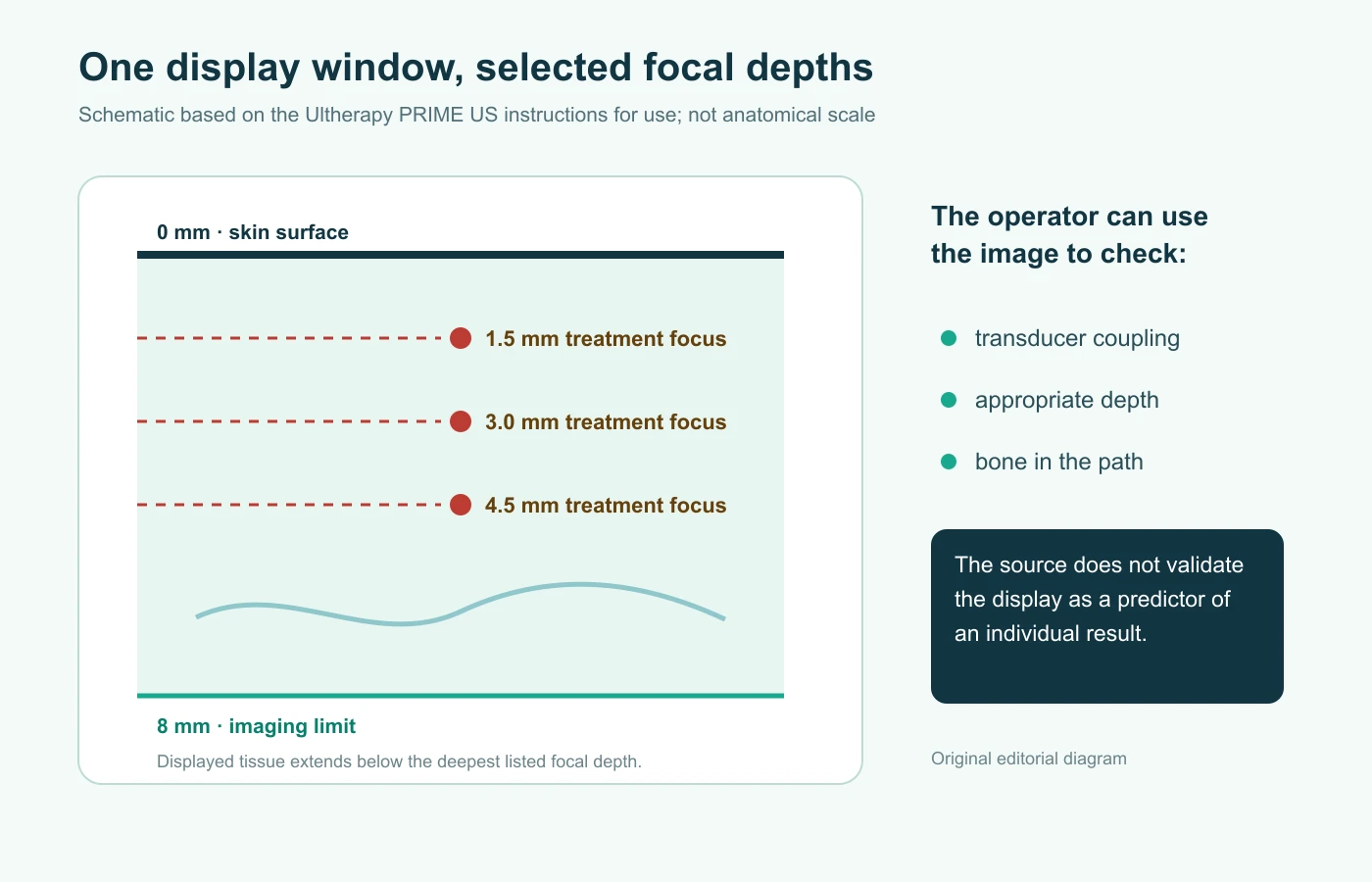
The current US Ultherapy PRIME instructions for use list an imaging depth of 0–8 mm for each of the six transducers. The same table lists treatment focal depths of 1.5, 3.0 or 4.5 mm, depending on the transducer. The instructions also summarize the system's treatment-depth range as 1.5–4.5 mm.
In other words, the 8 mm specification describes how much tissue can appear in the displayed field. It does not mean the system treats every layer from the surface to 8 mm. A treatment line creates discrete thermal coagulation points at the selected transducer's focal depth.
What the operator is checking
The US instructions state that the imaging feature lets the user view skin and subdermal regions before treatment. The listed purposes are to ensure proper coupling of the transducer to the skin and to confirm an appropriate treatment depth, with avoiding bone given as an example. The instructions tell the operator to place the treatment window flush with the skin, begin imaging and review the displayed tissue before delivering a line.
FDA's 2024 K233996 summary for the PRIME console repeats those functions. It also says PRIME and the earlier predicate use the same imaging mode. The earlier K180623 summary uses the same 8 mm range and the same coupling and depth language. Those documents support a procedural-control description; they are not a head-to-head test of outcomes with and without visualization.
What the image does not establish
None of these three records validates the display as a test for predicting how much lift a person will see, whether two sides will respond identically, how long a result will last or whether one console will produce a better clinical result. Imaging can make specific steps inspectable during the procedure without turning biological response into a certainty.
The labeled imaging uses are tissue visualization, skin coupling and confirmation of treatment depth.
Four questions to ask at a consultation
- Which system and transducer would be used for the area being considered?
- What will the operator check on the image before delivering energy?
- What finding would make the operator change position, depth or the plan?
- What can the image confirm during the procedure, and what remains uncertain about my result?
Ask the provider which system and transducers will be used. Imaging-guided treatment is described in Delight Dermatology's notes on Ultherapy Prime and DeepSEE.
Regulatory and geographic scope
The documents cited here are US manufacturer instructions and FDA 510(k) records. They do not establish Korean authorization, a provider's credentials or personal suitability for treatment. The screen is one input to a trained operator. It does not replace an examination, medical history or discussion of alternatives and adverse effects. For the broader mechanism and US regulatory history, see what Ultherapy is.